|
About Knee
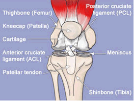
The knee is the largest joint in the body, and one of the most easily injured. It is made up of the lower end of the thighbone (femur), which rotates on the upper end of the shinbone (tibia), and the knee cap (patella), which slides in a groove on the end of the femur. The knee also contains large ligaments, which help control motion by connecting bones and by bracing the joint against abnormal types of motion. Another important structure, the meniscus, is a wedge of soft cartilage between the femur and tibia that serves to cushion the knee and helps it absorb shock during motion.
- The joint surfaces - The femur has 2 smooth, rounded joint surfaces. They move on the nearby flat joint surfaces on the tibia.
- The cartilages - To reduce friction, the joint surfaces are covered with cartilage. This cartilage is the smooth white layer that you see on the end of a lamb or chicken bone where it forms a joint. A "torn cartilage" does not refer to a problem with this layer.
- The meniscus - These are two half-moon shaped structures that sit on the top of the tibia. When people say that someone has a torn a cartilage in the knee they are actual¬ly referring to a torn meniscus.
- The patella - The patella is the bone at the front of the knee, commonly called the kneecap.
- Cruciate ligaments - These are two ligaments between the femur and the tibia. These ligaments make a cross, hence the name cruciate ligaments. One ligament starts at the front and is called the anterior cruciate ligament or ACL for short. The other starts at the back of the knee and is called the posterior cruciate ligament or PCL. These cruciate ligaments keep the femur and the tibia properly aligned as you bend and straighten your knee.

Common Knee Injuries
Anterior Cruciate Ligament Tear
 Anterior Cruciate ligament is torn while its ability to withstand the load is overcome by the force of injury. ACL is commonly injured with twisting injury to the knee, hyperextension injury and abnormal sideway opening of the joint ( varus or valgus). Depending on the amount of force, other structures are injured along with ACL. These include Meniscus, PCL and collateral ligaments. ACL injury is actually a spectrum of injury ranging from an isolated ACL injury (eg sports, low velocity injury) on one end to debilitating multiligament injuries ( Road accidents etc) on the other.
Like many other joint structures, ACL also has very poor blood supply and hence it has very poor healing potential. The healing potential is further compromised by the presence of joint fluid, which inhibits clot formation and healing response. Most of the time ACL is torn completely. Partial tears are rare and may progress to complete tears.
Clinical Features
When an ACL is torn the patient may feel a "pop" like sensation in the knee and may have difficulty in standing immediately after injury. In most of the cases, a knee swelling ( hemarthrosis ) develops within few minutes due to the bleeding from the torn ligament. However ACL can be torn without a knee swelling also. The subsequent disability of the patient depends on ACL deficiency as well as the amount of injury to the associated structures of knee. If only the ACL is torn, the patient will be able to walk, but the knee will buckle (give away) when he changes the direction while walking (pivoting and cutting). Additional injuries to meniscus and cartilage will result in episodes of locking, pain and joint swellings.
Diagnosis
Most of the ACL injuries can be diagnosed by clinical examination. Clinical diagnosis may be difficult if the patient is having lot of pain and spasm. In this situation an examination is performed at a later stage. MRI is valuable in diagnosing ACL as well as other injuries and is useful in some patients.
Treatment
Because of the poor blood supply, repair (bringing together the torn ends by suturing) will fail invariably and is no longer practised. Reconstruction of the ACL is the ideal treatment. In this method a band of tissue (Graft) is harvested from around the knee and is placed in the original position of the ligament. Two common grafts are used in our center.
The Bone patellar tendon bone graft (BPTB) graft consists of the middle third of the patella tendon, a small piece of bone from the knee cap and from the tibia .
Another graft source is the tendons of Hamstring muscles, semitendinosus alone or with gracilis. Allografts are tissues taken from dead donors. Though available in abroad, we do not use them currently.
For fixing the grafts, passages(tunnels) are made in the tibia and femur, the grafts are passed and fixed using implants. There are variety of fixation techniques and implants available.
Meniscus tears and management
 Meniscus is the shock absorber of the knee. Only the outer margins of this structure has blood supply and most of the inner regions lack any blood supply. Because of this only tears occurring in outer margins have the potential to heal. Meniscus tears occurs in various patterns. Meniscus tear can produce symptoms like pain, repeated joint swellings (effusions), locking and catching. Diagnosis of meniscus tears is by clinical examination and in doubtful cases, MRI.
Meniscus tears are treated according to the blood supply of the meniscus. Tears occurring in the regions with blood supply are treated with Meniscus repair. Those occurring the area without blood supply are trimmed to leave behind a smooth rim of meniscus. (Partial Meniscectomy). Early presentation for surgery is crucial for meniscus preservation. With late presentation, the chances of success following a repair come down because of poor tissue quality. Considering the vital role of the meniscus in knee function every attempt is made to preserve the meniscus. Meniscus is the shock absorber of the knee. Only the outer margins of this structure has blood supply and most of the inner regions lack any blood supply. Because of this only tears occurring in outer margins have the potential to heal. Meniscus tears occurs in various patterns. Meniscus tear can produce symptoms like pain, repeated joint swellings (effusions), locking and catching. Diagnosis of meniscus tears is by clinical examination and in doubtful cases, MRI.
Meniscus tears are treated according to the blood supply of the meniscus. Tears occurring in the regions with blood supply are treated with Meniscus repair. Those occurring the area without blood supply are trimmed to leave behind a smooth rim of meniscus. (Partial Meniscectomy). Early presentation for surgery is crucial for meniscus preservation. With late presentation, the chances of success following a repair come down because of poor tissue quality. Considering the vital role of the meniscus in knee function every attempt is made to preserve the meniscus.
Cartilage Injuries
 Cartilage is responsible for the smooth gliding movements of the joint. Cartilage injuries can occur along with ACL ruptures at the time of injury or because of the repeated instability associated with ACL deficient knee. Only a localized part of the cartilage is injured and hence they are called focal cartilage injury. Cartilage injuries are difficult to diagnose clinically and in MRI. Arthroscopy is the only modality, which can clearly visualize the injury and treat it. Cartilage is responsible for the smooth gliding movements of the joint. Cartilage injuries can occur along with ACL ruptures at the time of injury or because of the repeated instability associated with ACL deficient knee. Only a localized part of the cartilage is injured and hence they are called focal cartilage injury. Cartilage injuries are difficult to diagnose clinically and in MRI. Arthroscopy is the only modality, which can clearly visualize the injury and treat it.
The cartilage injury is graded according to depth and extent. Superficial lesions are debrided and smoothened. For full thickness lesions either Microfracture Chondroplasty or autologous osteochondral transfer (mosaic plasty ) is carried out.
Posterior Cruciate Ligament (PCL) Injury
Compared to ACL injuries, PCL injuries are rare and mostly missed. They occur in road traffic accidents and falls. PCL injuries cause the tibia to move backward abnormally. Once again the degree of PCL injury is graded according to the severity. Grade I and II are treated non-operatively. Grade III and IV injuries are treated by arthroscopic PCL reconstruction. For PCL reconstruction we use hamstring muscle tendon as graft.
Knee Dislocation
Knee dislocations are rare but can be devastating. They are associated with multi-ligament injuries. Following reduction and a period of rehabilitation, singe stage multi-ligament reconstruction is carried out to stabilize the knee.
Collateral Ligament Injuries
The Medial Collateral ligament which supports the inner aspect of knee is commonly injured. MCL injuries are graded according to the severity. Grade I being contusion, Grade II partial tear and Grade III being complete rupture.
MCL has a good healing potential and surgery is not required. MCL injuries are treated initially by rest, ice compresses, anti-inflammatory medications. Once the pain comes down the patient is ambulated with non-weight bearing and knee ROM.
The treatment of lateral collateral ( LCL ) injuries is much more complicated. Usually the LCL is injured along with the neighbouring structures called PLC ( Posterolateral complex). Most often this group of structures are injured along with ACL or PCL. As this complex is necessary for day to day function of knee, any laxity in these structures is unacceptable. Repair of the structures gives good results ( within 3 - 4 weeks ) and delayed cases need reconstruction of this structures. Good PLC function is important for the success of ACL or PCL reconstruction. Any PLC laxity found during ACL or PCL reconstruction is treated by reconstruction. PLC reconstruction has to be done by open appraoch only.
Patella Dislocation
The patella ( knee cap ) is also the source of many problems. The knee cap moves in a groove of the femur during knee flexion and extension. In some patients the knee cap slips out of the groove and comes to lie on the outer aspect of the knee. Many factors like, muscle imbalance, altered anatomical development and ligament laxity play a role in the causation of this problem. It requires surgical intervention if it occurs more than once. The surgical treatment includes strengthening the torn / loose ligaments and releasing the tight structures. In a select few, bony procedures to correct development bony problems may be necessary. The relevant procedures will be decided for each patient after thorough clinical and radiological evaluation.
Knee Pain
Knee pain is very common in young as well as old people. Not every knee pain is associated with structural damage. However, the cause of the knee pain has to be assessed clinically. If necessary, investigations like MRI are done. In the absence of structural damage to the knee, a rehabilitation programme is started with individualized physiotherapy to correct muscle imbalance and tightness.
Synovitis
Synovium is the lining membrane of the joint which secretes the joint fluid. The synovium can be affected in many diseases processes leading to swelling of the membrane, fluid collection. The basic disease producing the inflammation should be identified and treated. Rarely the synovium proliferates and produces cartilaginous nodules ( Synovial Osteochondromatosis). The nodules get detached from the synovium and become loose bodies in the joint. Arthroscopy is very useful in removing the loose bodies and the diseased synovium. Pigmented villonodular synovitis is another rare disease of knee producing painless swelling of knee and later joint erosions.
Arthroscopy has significant advantage in removing the synovium, when compared to open procedures. As the front, back and sides of the joints can be approached by few 10 mm skin incisions.
First aid in knee injuries
Look for obvious deformity, swelling, bony discontinuity.
- Feel the pulse in the foot and test for sensation in the foot.
- Support the limb with a splint. If proper splints are not available use board strip or a wooden slab to support the limb.
- Bleeding wounds - wash with clean water and cover with sterile dressings. Do not apply tourniquet / constricting bands
for stopping the bleeding.
- Rest to the affected part
- Elevate the part
- Ice packs - to reduce the pain and swelling.
- Gentle compression ( elastocrepe ) bandage.
- Medical opinion.
- MNEMONIC : PRICE ( Protect -Rest- Ice-Compression dressing- Elevation )
Treatment Options
- Not all knee problems require surgeries.
- Rehabilitation & physical therapy has a definite role in many problems.
- Arthroscopic surgeries can be performed on Day Care basis, Indoore basis.
Recovery
The recovery depends on what type of surgery is performed. One of the problems with knee arthroscopy is that the procedure hurts much less than open shoulder surgery, and therefore patients may tend to do too much, too soon. It is very important that you only perform activities that your surgeon recommends following a knee arthroscopy. Even though your knee may feel fine, you need to allow time for repaired tissues to adequately heal. This is especially important for patients who have cruciate ligament reconstruction & multiliagamentous repair and / reconstruction. Patients with small meniscus tear or loose body removal can resume their work day after surgery.
Physical Therapy
Physical therapy involves activities to restore your knee mobility, reduce pain and improve the strength of muscles around the joint. A physical therapist is a key part of our medical team, and will be involved in all phases of your knee rehabilitation. If your condition doesn't improve after a period of treatment, however, your physician may recommend surgery. Physical therapy will continue to be an integral part of your recovery after surgical treatment.
Advantages
Although arthroscopic surgery has received a lot of public attention because it is used to treat well-known athletes, it is an extremely valuable tool for all orthopaedic patients and is generally easier on the patient than "open" surgery. Most patients have their arthroscopic surgery as outpatients and are home several hours after the surgery.
Updates
- We are also doing mosaicplaty.
- Abrasion chondroplasty & microfracture chondroplasty.
|